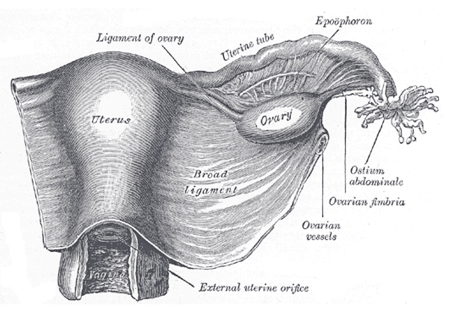
For more than a century, a small structure attached to the ovary was treated as an anatomical leftover with no real job. New research suggests that verdict may have been badly wrong.
The rete ovarii was first described in 1870 and appeared in illustrations in early editions of Gray's Anatomy, the landmark reference first published in London in 1858. Over time it vanished from medical textbooks and came to be regarded as a functionless remnant in the adult ovary.
That changed when a research team released a paper titled "Rediscovering the rete ovarii, a secreting auxiliary structure to the ovary," later published in the journal eLife, which examined how the structure develops and what it does in mice.
What Researchers Found
The rete ovarii is a horseshoe-shaped band of tissue at the base of the ovary. The team identified three distinct regions — the intraovarian rete, the extraovarian rete and the connecting rete — that persist from birth into adulthood.
When researchers injected fluid into the extraovarian portion, it traveled toward the ovary, hinting that the structure may feed into ovarian function. Its cells sit close to the blood vessels supplying the ovary, receive direct nerve connections and neighbor macrophages, the immune cells that help defend tissue.
The cells also carry receptors for estrogen and progesterone. Some of the proteins the structure releases are linked to egg maturation and to maintaining ovarian follicles, and that activity rose along with estrogen levels — suggesting the rete ovarii responds to the hormonal shifts of the reproductive cycle.
Co-author Dilara Anbarci, a developmental biologist at the University of Michigan, told Science News that the findings could shed light on fertility and ovarian health. "There's still so much we can't even begin to comprehend about female anatomy," she said.
The authors note that scientists have studied the structure on and off since it was first described but never reached agreement on its purpose. The new work was done in mice, and how closely the results translate to humans remains to be established.
A Viral Moment
The rediscovery has taken off on social media, where many women have pointed to it as an example of a historically male-dominated medical establishment overlooking female biology.
Comedian Michelle Wolf called the news "infuriating" in a TikTok that has drawn more than 130,000 likes, noting that the rete testis — generally considered the male counterpart — never left the textbooks, even though the study's authors describe the ovarian version as the more complex of the two. She closed by welcoming the rete ovarii back.
Based on reporting by the New York Post. Illustration: Uterus, ovary and uterine tube from Gray's Anatomy, 1918 edition (Henry Vandyke Carter, public domain).